

Take a Closer Look at Eye Exams
Bedside assessments provide key information about patient health and can serve as prevention tools for those at risk of falling.
Poor eyesight increases the risk of older adults falling during a hospital stay.1Eye problems are also a top reason for emergency department (ED) and primary-care visits.2Bedside eye exams can provide essential information about a patient’s health and serve as prevention tools for individuals at risk for falls. Common vision conditions that can increase the risk of falls include age-related macular degeneration, cataracts, diabetic retinopathy, and glaucoma.1
EYE EXAM PREPARATION
Hand-washing between patients is preferred over using alcohol-based hand sanitizer for eye exams,2and gloves can be worn for protection from blood, discharge, or tear secretions. Nurses can ensure that all necessary exam equipment is readily available, including bright light sources, hand-held magnifier, tissues, and topical anesthetics. It is also important to discuss what patients should expect during the eye exam.
EYE EXAM BASICS
The first step is to conduct a general inspection of the external eye by examining the eyebrows and checking for symmetry with expression changes, hair distribution, and skin texture.3This can provide insight regarding medical conditions. For example, unequal eyebrow symmetry can be the result of nerve damage, and short or thin eyebrows can indicate hypothyroidism. Next, the eyelid should be inspected for periorbital edema (eye swelling) or skin abnormalities. There are a variety of infections that can affect the superficial tissue surrounding the eyes, such as periorbital cellulitis. Other infections can include a hordeolum (sty) that involves an acute painful focal infection of the eyelid caused byStaphylococcus aureus, which is typically treated with a prescription ophthalmic antibiotic.3Other conditions that may be found upon exam include blepharitis (eyelash follicle inflammation) and a chalazion (eyelid swelling), because of blocked sebaceous glands.3It is important to report ocular problems to the patient’s health care provider for follow-up.
Examining the eyeballs to ensure they are aligned with their sockets and symmetrical is also an important part of the exam.3Exophthalmos (eyeball protrusion from the socket) can indicate Graves’ disease or thyroid dysfunction.3Check out the conjunctiva, the clear vascular membrane that covers the sclera, which should be clear with few or no red lines. Conjunctivitis, or “pink eye,” can be caused by allergies, bacteria, or viruses. The cornea can be examined by using a light source, and it should appear clear and smooth. If irregularities exist, there may be a corneal abrasion or laceration. More in-depth exams can be performed with a fluorescein stain and Wood lamp.3
Visual acuity can be assessed using the Rosenbaum Pocket Vision Screener Card for near vision, and the Snellen chart can be used for distance vision.3During bedside exams, these tests can evaluate patients for acute vision changes. Patients can be referred to an ophthalmologist or optometrist if their vision is not accurate. The confrontation test can be used to measure peripheral vision, which is conducted by the nurse standing 2 feet in front of the patient.3Instruct the patient to cover 1 eye while looking straight ahead as the nurse moves an object into the visual field and into a central position through many angles.3The patient then identifies when the object is viewed, and the results are recorded in degrees, with normal visual acuity as 50 degrees upward, 70 degrees down, and 90 degrees from each horizontal margin.3
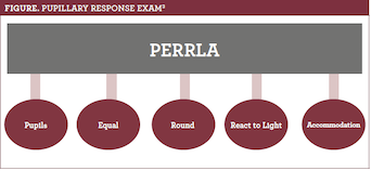
The charting abbreviation commonly known as PERRLA stands for pupils equal, round, react to light, and accommodation, and it is a quick 2-part test done in a dark room with a penlight (see figure).3During the first part of the exam, pupillary light reflexes are assessed. Constriction of the pupil that the light is shining in and simultaneous equal constriction of the other pupil is considered a normal finding. The second part of the exam tests for accommodation, which involves the patient focusing on a distant object and then looking at a near-central object. Normal findings when viewing at a distance are dilated pupils, and pupil constriction occurs while looking at near objects. The Glasgow Coma Scale, typically used in the ED to quickly evaluate a patient’s level of consciousness after a traumatic brain injury, also can assess eye-opening, motor, and verbal responses.3


BEDSIDE TOOL FOR FALL PREVENTION
There is a new bedside tool that provides nurses with a convenient way of assessing hospital patients at risk for falls.1,4This free resource, calledLook Out! Bedside Vision Check for Falls Prevention, was developed by the National Audit of Inpatient Falls in conjunction with British professional organizations, and it uses questions and visual aids for bedside eye exams.4Nurses can download this tool, which includes questions about patients’ eyesight, simple visual tests that check distance and near vision, and visual instructions to evaluate eye movements
and side vision.1,4
Jennifer Gershman, PharmD, CPh, is a drug information pharmacist and Pharmacy Times® contributor who resides in south Florida.
REFERENCES
- Windsor J, Dix A. A bedside tool to assess eyesight in hospital patients at risk of falls.Nursing Times. May 2, 2017. nursingtimes.net/7017618.article?search=https%3a%2f%2fwww.nursingtimes.net%2fsearcharticles%3fqsearch%3d1%26keywords%3dbedside+tool+to+assess+eyesight+in+patients+at+risk+of+falls. Accessed December 22, 2018.
- Marsden J. How to examine an eye.Nurs Stand. 2015;30(13):34-37. doi: 10.7748/ns.30.13.34.s44.
- Saathoff A. The nurse’s guide to bedside eye exams.Nursing Made Incredibly Easy. 2018;16(5):19-22. doi: 10.1097/01.NME.0000542480.68937.3d.
- Bedside vision checks for falls prevention: assessment tool. Royal College of Physicians website. rcplondon.ac.uk/projects/outputs/bedside-vision-check-falls-prevention-assessment-tool. Published January 23, 2017. Accessed December 22, 2018.